What is Macular Degeneration?

eye

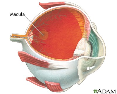
To understand age-related macular degeneration (AMD), the most common cause of legal blindness in older people, you need to understand the macula.  First, think of the eye as a hollow globe made of three layers.  The white, outside layer is the sclera. Inside the sclera is a middle layer called the choroid, which contains networks of tiny blood vessels called capillaries. The inner layer, called the retina, contains pigment-filled cells that absorb light, light receptor cells call rods and cones that change light into electrical information, and nerve cells that transmit that information to the brain. In the center of the retina, there is a tiny yellow spot about the diameter of a pencil eraser called the macula lutea (Latin for yellow spot).

What does the macula do? Try looking at a star to find out.

The macula contains only cone receptors, which detect color, and is responsible for sharply focused vision. If the macula degenerates, visual clarity is lost. The other 96% of the retina contains mostly rods, non-color receptors that perceive dim light and movement and are responsible for peripheral vision.  When you look at stars, you see them only with your peripheral vision. Trying to focus on a single star makes it disappear because the macular cones require more light than the night sky offers.  Imagine the disappearance of anything you try to focus on, in any kind of light, and you have some grasp of what macular degeneration is like.

When the macula degenerates

Age-related macular degeneration moves slowly, with many years between the first visible changes in the retina and the onset of symptoms. The first symptoms come from decreasing visual sharpness. Reading speed declines. Reading glasses no longer work for fine print. Road signs look blurred. With progression, dark patches or blank spots appear when patients focus on faces or print, and sometimes straight lines appear wavy. Legal blindness (best corrected vision less than 20/200) comes later and is never total because peripheral vision is spared, even though some of the same degenerative changes affect other parts of the retina. The macula is simply so tiny and so specialized that small areas of damage interfere with the ability to see clearly.

What’s the cause of macular degeneration?

No one knows for certain what causes macular degeneration but ophthalmologists suspect that the vascular anatomy of the retina plays a significant role. While the macula looks yellow, the rest of the retina has a reddish hue because of an overlying bed of capillaries, branches of the retinal arteries and veins which spread like a tree over the retinal surface. The tree stops at the macula and this vital region depends solely on blood flow through the network of tiny capillaries in the choroid layer for oxygen and nutrients and for clearing away the products of its high rate of energy use. The first changes that signal possible AMD, visible long before any symptoms appear, occur in the microscopic space between the choroid layer and the retina and are called Drüsen.

Signs of deterioration

Drüsen are yellowish accumulations of fats and proteins and inflammatory substances.  Small, sharper edged patches called hard drüsen are commonly seen in other parts of the retina and do not interfere with vision, but larger, fluffy patches, especially in the area of the macula warrant more frequent follow-up exams because people who have them sometimes, but not always, develop symptoms of AMD.  If visual symptoms occur and drüsen are present, the diagnosis is dry macular degeneration. In about 10-20% of cases of dry macular degeneration, new tiny blood vessels poke through to the retinal layer from the choroid, a process called neovascularization. When this happens, the diagnosis becomes wet macular degeneration which reflects the tendency of these blood vessels to leak and bleed, causing more cell destruction and separation of the retina form its underlying supporting layers.

Who’s at risk for macular degeneration?

The primary risk factors for macular degeneration are age, smoking and increased body mass index. A family history of macular degeneration also increases the chances of its occurrence, but so many different genes are involved that predictive genetic testing is not useful. Diabetes and vascular disease can accelerate the degenerative process. On the positive side, diets rich in green leafy vegetables, particularly those containing abundant antioxidants called carotenoids, as well as diets rich in Vitamins C and E, and those rich in zinc have been associated with lower frequencies of macular degeneration.

Other eye problems are more common

Fortunately, AMD is much less common than other age-related visual problems like presbyopia (poor near vision) and cataracts, both of which are eminently treatable. AMD affects about 2% of people in their 70s. Prevalence jumps to about 14% in Caucasians who reach their 90s, but remains at about 2% in other races. Legal blindness typically does not occur until the eighties.

Treatment

There is no treatment for dry macular degeneration.  The positive correlation between dietary antioxidant consumption and lower rates of AMD has led to the development of the “eye vitamins,” and while studies have shown some slowing of the degenerative process when these vitamins are consumed, there is no evidence that taking them prevents the onset of the disease.

The first line of treatment for wet macular degeneration is injection, directly into the eye, of drugs which block new blood vessel formation. This slows the disease process, but does not cure it. Another treatment is photocoagulation, or the injection of drugs which, once activated by light aimed at the new blood vessels causes them to shrivel.  Laser treatment of troublesome blood vessels is commonly done, but it destroys the retina in the area treated, so its aim is prevention of new problems.  Surgery is sometimes required to drain fluid accumulations or reattach retinal tears.

Preliminary studies suggest that high dose cholesterol lowering drugs (statins) may shrink drüsen, but one worrisome study of data from a large managed care group suggested that statin use for over a year increased rates of progression of dry AMD to wet AMD. Long term prospective studies are needed, and eye exams should be routine for anyone taking statins.

Useful Macular Degeneration websites

https://nei.nih.gov/health/maculardegen/armd_facts

Home Page

http://www.aao.org/eye-care-for-older-adults

http://www.amd.org/

Broken Heart Syndrome: The Octopus Trap

“Doctoring her seemed to her as absurd as putting together the pieces of a broken vase. Her heart was broken. Why would they try to cure her with pills and powders?”  Leo Tolstoy, writing about Kitty’s heartbreak over Vronsky in Anna Karenina


Sometimes people say that a spouse who dies unexpectedly within hours to weeks after the partner’s death has “died of a broken heart,” though a variety of different medical conditions are responsible for the increased death rate among grieving partners, who are often elderly. In 1990 a paper appeared in the Japanese medical literature that described a peculiar heart problem, documented by modern technology, that the popular press seized upon as a possible explanation for the correlation between grief or fright or other emotional stress and sudden, unexpected death. The cardiomyopathy the authors described was an abnormality in the heart muscle of the left ventricle, the chamber of the heart that pumps blood out to the body. That part of the heart acted as if it had been “stunned” into inactivity and caused pain and other symptoms commonly associated with heart attacks, but the patients did not have any coronary artery disease.  These facts seemed fit neatly into the concept of a “broken heart.”

Why an octopus trap?

The ventriculograms, or dye studies, of the hearts of the Japanese patients described in the 1990 paper showed peculiarly dilated left ventricles, ballooned at their tips so that they resembled octopus traps – narrow-necked, flask-shaped contraptions that are easy for the tentacled animals to enter but hard to escape. In the Japanese language an octopus trap is a takot-subo and by the mid-2000s the name Takotsubo cardiomyopathy, or TCM, was widespread and many more cases had been described. Risk factors for the stress-induced cardiomyopathy were both physical and mental and included stays in ICUs, near drownings, major physical injuries, bad medical or financial news, legal problems and natural disasters, and, of course, unexpected death of a loved one. Cases have also been attributed to cocaine and methamphetamine use, as well as to exercise stress testing. These patients who acted as if they had had a heart attack were most often women and they had no history of heart problems prior to the events that hospitalized them.

Who is at risk? What are the symptoms?

Takotsubo syndrome is not common, but also not rare. It accounts for 1-2% people who have symptoms initially thought to be caused by regular coronary artery disease. In women, some people estimate that as many as 5% of heart attacks are actually TCM.  Most TCM patients are Asians or Caucasian, over 90% are post-menopausal women and most cases come to attention because of heart attack-like symptoms such as acute chest pain and shortness of breath.  But unusual presentations also occur as a result of the effects of the poor heart muscle function. When it’s pump action fails, the heart sends hormonal signals that affect water and salt balance in the body.  Fluid retention occurs in some people. Low sodium levels cause symptoms of profound fatigue in others. Clots may form in the poorly contracting ventricle, break loose and cause strokes. Lethal complications such as ventricular fibrillation and actual rupture of the impaired ventricle are very rare, but have occurred.

What’s the cause?

Diagnosis of Takotsubo syndrome requires new abnormalities in the electrocardiogram, absence of coronary artery disease and no evidence of heart inflammation from an infection or autoimmune disease. While the enzyme markers for a heart attack may rise, they do so earlier and fall back to normal more quickly than they do in a routine heart attack. In addition, the muscle abnormalities in the left ventricle can’t be mapped to the territory supplied by one coronary artery as they can when a blockage is responsible for the damage. Doctors who make a TCM diagnosis must also make certain the patient does not have a tumor called a pheochromocytoma, which produces stress hormones.

Most patients recover completely

By now TCM is known to be transient, with supportive care leading to complete recovery within 1-2 months in over 95%of patients. Recurrence is extremely rare. However, the actual cause, or mechanism by which the transient heart damage occurs, remains unknown. A number of theories have been proposed and all of them have something to do with a temporary derangement in function of the cells of the inside layer of cells of the left ventricular chamber of the heart. In these cells normal energy production from fatty acids is halted. The area of the heart involved happens to have a high concentration of receptors for catecholamines (adrenaline like hormones), perhaps making it susceptible to overstimulation and damage by severe stress. The high preponderance of postmenopausal women in case reports suggests that perhaps sex hormones are somehow protective factors.

Do people really die from broken hearts?

But is the Takotsubo syndrome responsible for deaths that seem to come from emotionally broken hearts? The mortality rate in cases of Takotsubo syndrome that come to medical attention is low. Recovery rates are high. Broken heart deaths most often occur in older people who have multiple health problems which might play a role. For example, when singer/actress Debbie Fisher died as she was planning her daughter Carrie Fisher’s funeral this year, a NYT reporter speculated about the cause of death being the Takotsubo syndrome. But Debbie Fisher had suffered several strokes in recent years and had high blood pressure. Later stories attributed her death to a fatal stroke related to high blood pressure.

Grief and stress do raise the risks of dying for the bereaved, but the causes of death are many and varied and mostly related to longstanding health problems.  The pills and powders Kitty scorned for her broken heart in Anna Karenina have a place in the treatment of the many other problems that occur in the setting of grief, especially depression. While it is tempting to attribute sudden, unexpected deaths in emotionally stressed people to an odd and mysterious heart problem named after an octopus trap, science requires objectivity and evidence.  So far the evidence about sick hearts that resemble octopus traps suggests that, at least in the people in whom the diagnosis is made, death is a very rare outcome and complete recovery is the rule.

Chilly Treatment

Large scale studies of survival after cardiac arrest have produced dismal statistics, with survival to hospital discharge of 17.6% when the patient is already in the hospital at the time of the arrest and only 6.1% when the arrest occurs outside the hospital.  The development and widespread deployment of portable automatic external defibrillators (AEDS) in public places has increased the number of people who make it to the hospital after cardiac arrest.  However, the survivor’s longer term outcome depends in large part on how much brain damage occurs during the arrest and whether or not the restoration of circulation damages the brain further, a phenomenon called reperfusion injury.  Because the odds of initial survival have improved, and because lowering the core temperature of the body appears to lessen reperfusion injury, the subject of hypothermia has emerged as a vibrant area of research and therapeutics.

Therapeutic hypothermia is an old idea

Hippocrates (460-370B.C.) recognized the value of cold temperatures in the outcome of soldiers with head injuries and in people suffering from tetanus. In the 1800s, Napoleon’s surgeon used ice to prepare limbs for amputation because it numbed pain and reduced bleeding. Over much of history, miraculous recoveries were reported in victims of cold water submersion. But not until the late 1950s did therapeutic hypothermia become a routine part of some surgical care, when experiments in animals demonstrated its value in protecting the brain during the open-heart surgery.  Despite some attempts to cool patients for other problems such as cardiac arrest, strokes and head injuries, the number of problems encountered in during cooling and in the re-warming phase put a damper on the use of the technique. Now, however, we are in the middle of a revival of interest in therapeutic hypothermia.

How does cold help?

Cold protects the brain because the biochemical reactions that sustain life are influenced by temperature. If the heart stops, the brain runs out of fresh supplies for energy production in two minutes. A downward spiral toward brain cell death begins unless blood flow is restored within the next two minutes. When blood flow is restored (reperfusion), a cascade of potentially damaging chemical reactions begins in cells that have been deprived of oxygen. The longer the period of arrested circulation was, the more damaging these reactions are.

The body at different temperatures

Changing the body’s temperature changes the speed and efficiency of its chemical reactions.  At temperatures over 105 many processes fail completely. As body temperature falls below normal, chemical reactions slow down.  Between 92 and 89.6  the damaging chemical responses that come after blood flow returns are blunted enough to improve outcomes significantly.  By 90, pulse and respirations slow and peripheral circulation shuts down. By 86 the patient may still be alive, but looks dead. This level of deep hypothermia is used for some long, difficult cardiac and neurosurgical procedures.

Lowering temperature is now routine, sometimes

Since 2005, the American Heart Association has recommended therapeutic hypothermia as a routine part of patient care after a cardiac arrest in circumstances that depend on the reason for the cardiac arrest, the speed of the resuscitation, and the state of the patient after circulation is restored.  Some medical centers are also experimenting with the technique in the treatment of certain types of strokes and head injuries.

How do you lower someone’s core temperature?

How do you cool a body that normally maintains a constant temperature that hovers within a few tenths of a degree of 98.6?  Any environment with a temperature less than body temperature provides a gradient for heat loss.  Deliberately making someone hypothermic means increasing that temperature gradient. In the presence of a gradient, heat radiates away from the body. Heat is also conducted away when the body is in contact with any colder substance; when the colder substance such as air or water is in motion, heat is lost even faster, by convection.  Heat is also drawn away by evaporation of perspiration on the skin’s surface, where the sweat keeps the microclimate humidity at 70% even when you think you are dry.   Heat also dissipates when warm moist air is exhaled from the lungs.

Several internal and external ways of changing the temperature gradient  exist: ice packs applied to the head, neck, axillae, groin, where large blood vessels are close to the surface; cooling blankets that house cold water circuits; closed catheters through which cold saline circulates inserted into large blood vessels; ice water balloons in the bladder.    These methods are directed at the core temperature of the body – the temperature of the internal organs and the brain. They do not cause problems like frostbite, seen commonly with accidental hypothermia, because the ambient temperature is not freezing and the skin is protected from direct exposure to ice packs being used for cooling.

The body resists lowering the temperature

Normally, we protect ourselves from falling temperatures by putting on more clothes and increasing activity, and by shaking and shivering, which produce heat.  A patient who has suffered a cardiac arrest will not engage in the normal behavioral responses, but he will shiver and perhaps become agitated, both of which are counterproductive to getting the temperature down. Sedation and even muscle paralysis are therefore necessary for the period of cooling.

Despite problems,  therapeutic hypothermia is here to stay

Current therapeutic hypothermia protocols call for maintenance of temperature between 32-34 (89.6-93.2) for 18-24 hours, followed by passive re-warming over the next 24 hours. Overshooting and undershooting of temperature are both common, as are difficulties maintaining electrolyte and sugar balance. Some complications like pneumonia and bleeding problems are more common than in similar patients not being treated with cold temperatures. Much work remains to determine the best timing for induction and maintenance of hypothermia after cardiac arrest, but it is clear that “the sooner the better” is the general rule and that the revival of interest in therapeutic hypothermia is here to stay.

Tetanus: Poster Child for Preventive Medicine


True preventive medicine is an intervention that stops a disease from developing, not one which simply slows disease progress. The body’s immune system is the master of disease prevention and it is no accident that one of the first medical efforts at preventing disease stemmed from the observation in the late 1700s that suffering a mild infection like cowpox prevented a similar but more severe infection – smallpox. Immunization was born, and to this date is the single most effective form of prevention of lethal disease. In the current age of rejection of routine immunization by a significant number of people, the disease called tetanus and its prevention by immunization is a story worth reviewing.

What causes tetanus?

Tetanus a disease is caused by a type of bacteria called Clostridia tetani, a fragile little organism that can’t tolerate oxygen or high temperatures but which changes itself into a tough intermediate form called a spore to lie in wait for potential victims. C. tetani spores survive indefinitely, are common in soil, particularly manure rich soil, and are found in intestinal tracts of farm animal, cats, guinea pigs, rats and people. They can survive oxygen rich environments, the usual antiseptics and even the temperatures used to sterilize medical instruments. Once the spores gain entry into body tissues, they revert to fragile bacterial form, reproduce and begin to manufacture tetanospasmin, one of the most lethal toxins known to man and the substance responsible for the symptoms of the disease. Though farm animals and people are susceptible to tetanus infection, dogs and cats are not.

Development of symptoms

Tetanus infections are usually acquired when C. tetani spores enter the body through a deep wound in the skin that air does not reach. Contaminated batches of heroin are also sources of infection when the drug is injected under the skin or intravenously. In the first few days after C. tetani spores come to life inside the body, no symptoms or tests indicate anything amiss. As the toxin producing bacteria increase in number, and the toxin produced finds its way to the spaces between nerves and muscle and between motor nerve cells in the brain and spinal cord, profound muscle spasms begin. Tetanospasmin works by the blocking normal neurochemical signals that inhibit muscle tone and motor nerve excitability.
The time from infection to development of symptoms in any infection is known as the incubation period. In human tetanus, the closer the entrance point of the bacteria to the brain or spinal cord the shorter the incubation period. On average symptoms begin about a week after injury. Though localized forms of tetanus can occur, with muscle spasm limited to the area around the wound, most cases are general and symptoms begin in the muscles of the head and neck. Spasm of the powerful masseter muscles of the jaw is the origin of the term “lockjaw,” a commonly used name for tetanus infection. Vocal cord and respiratory muscle involvement can interfere with breathing. Abdominal, trunk and skeletal muscle involvement are extremely painful and spasms can be strong enough to fracture long bones and spinal vertebrae. Other complications arise from involvement of the central nervous system: fever, high blood pressure, heart rhythm abnormalities and seizures. Secondary complications like bladder infections, pneumonia and blood clots in the legs and lungs also contribute to the lethality of the disease. In the pre-immunization era, treatment was confined to supporting the patient through the four weeks it takes for the toxin’s effects to wane.

Making the immune system remember the disease

Unlike cow pox, in which the natural immune response directed against the cowpox virus prevents more cowpox episodes but also smallpox, a full-blown case of tetanus does not confer any immunity because the minute amounts of toxin that produce the symptoms are not sufficient to stimulate the immune system to make antibodies against it. Immunization to tetanus is accomplished by presenting the immune system with a much larger amount of a formaldehyde weakened version of the toxin, to which it will produce antibodies which will neutralize the real toxin should it ever appear. This process takes a few weeks and several doses are required over time to reach full potency of an antibody response.

Immunization programs have made tetanus rare enough for people to forget how terrible an illness it is. In the US, since routine, active immunization began in the 1940s, tetanus rates declined steadily and were at an all-time low of .01 cases per 100,000 people in 2009. In addition, with better supportive care, mortality rates declined from 30% in the mid-1900s to 10% in the first decade of the 21st C.

Borrowing someone else’s immunity

Nevertheless, tetanus infections still occur and may increase in frequency if immunization rates drop. Fortunately, another type of immunization helps when tetanus develops in people who have not been immunized – a passive immunization process that allows patients to borrow antibodies produced in the blood of other people who have been immunized against C.tetani. This “antitoxin” is a mixture of human gamma globulin from screened donors and antibodies in it that “recognize” tetanus toxin react with the toxin circulating in the tetanus victim’s body, neutralizing a lot of its potency.

Boosting weakened immunity

The antibody response to tetanus toxoid wanes over time, but a repeat injection brings it up to full speed quickly. Booster doses are recommended for all adults every 10 years and in the event of penetrating wounds, especially if immunization status is unknown. Awareness of the symptoms of tetanus and the status of immunization of anyone someone suffering from heroin addiction, a sad and growing problem, is crucial for anyone who cares for them. Tetanus is the poster child for preventive medicine and no one should have to suffer this disease. The earlier it is recognized, the better the outcome is likely to be.

Colds and Flu: Variations on a Theme

    You wake up one morning with a scratchy tickle at the back of your throat. It’s nothing, you think, but by the evening you’re worried. What are you coming down with now? In all likelihood, it’s just a cold. The sore throat will persist for a few days, along with a runny nose and stuffy head. You may develop a cough. About a week after the scratchy throat began you begin to forget about the cold and by ten days it is a memory. Your body has met a small bit of protein-coated RNA called a virus, and defeated it. 

The difference between a cold symptoms and flu symptoms

    The next month another bit of protein-coated RNA called an orthomyxovirus finds its way into your nose. This time the story is very different. At noon, you are feeling well. By 4PM you have been felled. Getting up from your desk feels like climbing Mount Everest. You cannot get warm – even under a pile of blankets. Then come the muscle aches, as if you’d run a marathon. This time there is no hope of sticking to a routine – you have the flu. The worst of the symptoms fade after a week, but fatigue and lack of energy may persist for several more. Rest, time and patience eventually lead you back to health. Next year, you think, you will get that flu shot. 

The viruses that cause the problems

    The viruses that caused these two illnesses are superficially similar. They are little protein bags full of bits of RNA– technically not living things, but able to commandeer the machinery of living cells to reproduce themselves. Carried in droplets spewed into the air by coughs and sneezes of infected people, they are directly inhaled or carried into the nose and mouth on fingers from surfaces where those droplets landed. Specialized proteins on viral surfaces attach them to the cells in the upper airways. These proteins (H, or hemagglutinin and N, or neuraminidase) and their numbered subtypes (1, 2, or3) give flu viruses their unimaginative names. Swine flu virus becomes H1N1.   

    For reasons not yet understood, the body’s response to the attack of cold viruses is not as severe as it is to flu viruses. Once inside cells, flu viruses elicit a flood of proteins called interferons, which are the source of fever, muscle aches and profound fatigue. The viruses rapidly go about the business of making more of themselves, sending them out to infect more cells and generating a new wave of symptoms. In the meantime, white blood cells begin to produce antibodies that target viral surface proteins and prevent them from locking onto more cells. Eventually the tide of the battle turns and no more cells are infected. The rest of the illness -the recovery phase – involves cleanup of the remnants of the fight. 

Why flu shots don’t always work
    Flu shots use mixes of several different flu virus types to stimulate the body to make antibodies to those viral surface proteins in advance of “catching” the flu. If and when the virus invades, the antibodies are in place, ready to block the initial attachment to cells in the upper airways. Because the flu viruses that travel the world vary over time, each year’s flu vaccine is a composite of some of the currently circulating strains and may or may not be a good match to the virus that ultimately shows up.  

    Immunization is recommended for children (older than 6 months), the elderly, people in the health and teaching professions, those living in nursing homes and dormitories, and for all age groups who are at risk for flu complications, namely those with other respiratory problems, cardiac disease, diabetes and immune system impairments (including those induced by treatment of other diseases like cancer). Most adults in good health have the immune wherewithal to recover from flu and colds without complications or medical intervention. 

Complications of the flu

    The complications of the flu and of colds are similar. Both infections affect the upper airways, causing swelling and inflammation of the linings of the nose, throat and sinuses and blocking narrow passageways within. Earaches and pain in the forehead or the face develop but often resolve with simple measures like propping head up while sleeping, and taking an anti-inflammatory medication like aspirin or Advil. Unremitting pain, with or without fever, may indicate secondary bacterial infection of the ears or sinuses – the only reason for the prescription of an antibiotic during a cold. 

    Pneumonia is the most serious flu complication. Pneumonia means that the lungs’ spongy structures where carbon dioxide is exchanged for oxygen are swollen and filled with inflammatory debris. Reappearance of fever and fatigue after the flu has begun to improve may be the first sign of pneumonia. Other symptoms are chills, chest pain, shortness of breath, and dry or productive cough. Sometimes the pneumonia is caused by bacteria, but more often by the original virus or another. Pneumonia caused by bacteria, at least outside hospitals, is much less common. Prompt medical attention is in order because oxygen levels may be low. Medical attention should also be sought for other delayed symptoms like change in mental status, particularly in babies and the elderly. Occasionally diarrhea and vomiting are part of the flu and may produce dehydration.       

Why hydration is important

     Dehydration is also a result of fever, the metabolic equivalent of exercising in a warm environment. Dehydration stresses already inflamed airways. The easiest way to keep track of hydration status is to look at your urine – the darker the color, the drier you are. Aim for almost clear urine by drinking plenty of water.  

Managing fever

    Fever, while distressing, is part of the body’s defense against cold and flu viruses, which thrive in the relatively cooler temperatures of the upper airways. Some of the fever related discomfort can be relieved with a bath. Aspirin is not to be used in children with the flu because of a rare complication called Reyes syndrome, a liver failure. Tylenol is safe for them.

  What about anti-viral drugs for the flu

     Antiviral drugs (Tamiflu, Relenza. Symmetrel, and Flumadine) drugs are only effective if begun in the first 24-36 hours after symptoms begin. They lessen the duration of symptoms by about a day. Widespread use will produce increasing numbers of drug resistant strains of viruses.  

    New flu viruses appear regularly and prompt anxious comparisons to devastating epidemics of the past. Worrying about them doesn’t help. Taking care of what you can do for yourself and your contacts (see below) is your best option. 
                                                                      Useful actions to prevent colds and flu

  • Maintain good health habits throughout the year: adequate sleep and exercise, nutritious diet, no smoking, modest alcohol use.
  • Cover your nose and mouth with a disposable tissue when you cough or sneeze, with the crook of your elbow in the absence of a tissue.
  • Wash your hands with soap and water or an alcohol based cleanser after coughing or sneezing, before eating, and after being out in public.
  • Keep your hands away from your eyes, nose and mouth.
  • During flu outbreaks avoid crowded, closed environments when possible and wash hands when you come home.
  • Remember that your flu is infectious for up to 7 days and try to avoid infecting others.
  • Get a flu shot if you are in a high risk group.

Heart Failure: The Price of Success

In 1953, surgeon John Gibbon undertook the first successful open heart surgery using a heart-lung machine, a pump that performs both heart and lung functions while the heart is stopped for repairs.  The pump ushered in a new era of cardiac surgery and made it possible to correct heart problems that had up until then caused premature disability and death.

In the years that followed Dr. Gibbon’s groundbreaking surgery, materials science, technology and pharmacology advanced rapidly, allowing surgery and medication to be used to treat a wider variety of heart problems, including heart attacks. Today we live in an age of coronary artery bypass surgery, clot-busting drugs,  and stents that prop open diseased coronary arteries. Advancements such as these have reduced death rates from heart attacks substantially; nonetheless, heart disease remains the number one killer of men and women in the United States, and heart failurethe constellation of symptoms that come from poorly contracting heart muscle –  is now the leading cause of hospitalization for patients over the age of sixty-five. With all of the progress that has been made in cardiovascular care, how can this be?

Why is there more heart failure now than in the past?

The answer is simple. Mortality rates are lower in the immediate aftermath of heart attacks because of the ability to dissolve clots and prop arteries open. Drugs and lifestyle management may be slowing the progression of the coronary artery disease that causes heart attacks. As a result of these advances, people no longer die as often in the earlier phases of a very long disease process which often ends in failure of the heart muscle to contract as strongly as it needs to.

If you liken cardiovascular care to home repairs, then the major advances in care ­­­have taken place in areas roughly equivalent to plumbing and electrical maintenance. Angioplasties, coronary artery bypasses, and stents are used to keep the pipes open; electronic pacemakers provide power, triggering muscle contractions in an organized fashion, while defibrillators restart the power when there are outages. A house with functional plumbing and electrical systems remains habitable remains habitable long enough for the underlying structural elements of the building, like the roof, ceilings, walls, and floors, to begin to fail. In the heart, the underlying structural element is muscle. When the muscular structure of the heart begins to fail, the signs and symptoms of heart failure appear.

Signs and symptoms

Heart muscle fibers that begin to contract less efficiently reduce the heart’s pumping capacity, preventing adequate blood flow to the major organs. The body reacts to this deficient blood flow by increasing the volume of blood in the body. It accomplishes this by retaining more salt and water, but when this happens, the blood’s increased volume stretches the failing heart muscle and damages it even further. The more the heart stretches, the less efficiently it pumps; the less efficiently it pumps, the more the body tries to boost blood flow by retaining fluids.

This vicious cycle results in what is known as congestion. Congestion occurs when the tissues, including the lungs, becoming filled with excess fluid. This fluid buildup causes swelling in the legs and abdomen and a shortness of breath. Fatigue and an inability to tolerate exercise are the heart failure patient’s constant companions. Gravity causes fluid to collect in the lower half of the body while the patient is standing or sitting, and when he lies down, it becomes redistributed, accumulating in the lungs where it can cause a telltale shortness of breath that is symptomatic of a deteriorating heart.

Causes

Age is an important contributing factor in the development of heart failure because the longer people live, the more time there is for coronary artery disease and the problems that contribute to it to take their toll on the heart muscle. Coronary artery disease deprives areas of the heart of blood and oxygen, leaving behind damaged muscle that contracts poorly and moves blood inefficiently. Smoking, excessive alcohol use, diabetes, hypertension, obesity, and lack of exercise contribute not only to coronary artery disease, but also to weakening the heart muscle directly. Less common factors involved in heart failure include viral infections that affect the heart and a variety of rare metabolic conditions that disrupt heart muscle fibers. Heart valve disease, when left untreated, can ultimately damage the heart by dilating it or causing it to thicken. However, valve disease is much less prevalent since the development of successful antibiotic treatments for childhood streptococcus infections.

Treatment

The quality and length of life heart failure patients can expect depends on how closely they adhere to the treatment plans provided by their doctors. Salt-restricted diets are a very important part of a heart failure treatment plan, and the mainstays of drug treatment plans are medications that prevent salt retention, get rid of excess water, improve the ability of the heart muscle to contract, and decrease blood pressure. Heart failure treatment treads a fine line between causing the patient’s body to retain too much and too little fluid. Drying a heart failure patient out too much can push the kidneys into failure. Too little and the lungs are liked soaked sponges, unable to exchange carbon dioxide for oxygen efficiently.  The margin of error in fluid balance gets smaller and smaller as the disease progresses and this tightening window contributes to high hospital readmission rates for congestive heart failure patients.  Physicians must monitor their patients’ weight, symptoms, electrolytes, and kidney functions more and more closely and start adding tests like chest X-rays and echocardiograms.

A variety of innovative devices and surgical procedures have been designed to cinch up dilated, failing hearts but have not succeeded in producing adequate results. Some success is being reported from the use of multiple pacemakers, which allow different segments of the heart to be stimulated in a defined order that improves the sequence of muscular contraction enough to generate greater cardiac output. Currently, bi-ventricular pacing—the separate pacing of both sides of the heart–is the most promising addition to the heart failure treatment arsenal. Cardiac transplantation remains the most difficult, expensive, and uncommon solution for a heart that has reached the end of its functional life.

Heart failure may eventually be overcome by artificial pumping devices or methods of stimulating the production of new cardiac muscle, but in the meantime, prevention is still the most desirable treatment option. Not smoking, maintaining a healthy weight and good exercise habits, sticking to a balanced diet of fresh foods, getting adequate sleep, and managing stress well are all cheap and valuable ways to invest in your heart’s health.


                          A Balanced Life

Humpty Dumpty sat on a wall,


Humpty Dumpty had a great fall. 


All the king’s horses, 
and all the king’s men,

Couldn’t put Humpty together again.

No one ever said why Humpty fell off the wall. If he’d managed to stay up there, he would have been OK. That’s the way it is with older people with thin bones. Osteoporosis doesn’t make people fall but it makes them break when they do. The real question is why they fall.

Why do older people fall down?
Falling is a risk of age because balance, strength, flexibility and speed decline over time. Even if you have no problems with balance it is worth understanding how balance works – how you maintain an upright posture and adjust to changes in the terrain under your feet, and how you manage to catch yourself and not fall as your foot slips on an icy path. The good news is that “use it or lose it” applies to balance, strength, flexibility and speed. You have some say in their preservation.

The systems that create balance

Your sense of balance comes from the integration of messages from muscles and joints, eyes and ears. Try experimenting and you’ll feel how these sensations contribute to balance. First, stand on one leg. Then try doing it with your eyes closed. Then try doing it after spinning around in a circle, and disturbing the fluid in the inner ear. With each maneuver, you subtract some of the sensory input to your brain and make it harder to control the muscle strength and tone needed to keep you upright. Fortunately, we don’t have to “think” about the actions that keep us balanced. They happen automatically.

What happens when the balance systems go awry?

When some part of the entire balance system goes awry, you feel “dizzy” or “lightheaded” or “off” or “tipsy.” The doctor who hears your complaint will ask you questions related to all the components of the balance system, and to all the medical conditions that can disrupt your eyes and ears, your peripheral nerves, your spinal cord, or your brain. He or she may order hearing tests or brain scans, blood tests or electroencephalograms. Patience, careful observation of symptoms, and systematic ruling out of problems is the best approach.

Vertigo

A sense of spinning dizziness, called vertigo, makes balance almost impossible. Vertigo is most frequently the result of an inner ear problem Three semicircular canals deep in each ear lie at right angles to each other and are filled with fluid that moves when you move your head. The fluid stimulates nerves that add information to the balance system. Viruses can affect the ear and produce profound vertigo with even tiny head movements. Some tumors of the nerve to the ear (acoustic neuromas) affect balance and hearing. A benign condition called Meniere’s disease causes episodes of hearing loss and vertigo. Though acute ear problems are sometimes at fault, very often dizziness that comes from the ears is a result of disuse of the inner ear canals. Ears that are unaccustomed to change in position because body movement has become limited and slow no longer cope well with rolling over in bed or turning the head quickly, and such routine activities can make the room spin. This is called benign positional vertigo and the treatment consists of exercises of the head and neck to re-accustom the semicircular canals to movement.

Muscle and joint receptors keep track of the body in three dimensions

Tiny receptors in the muscles and the joints perceive gravitational stress and muscle tension and movement. These receptors tell the brain where the body is and how much muscle tension is needed to hold you up and to move the way you intend to move. Balance suffers when nerves don’t function properly (neuropathies) because of diabetes, kidney disease, vitamin deficiencies, medications, exposure to toxic substances, or a variety of esoteric blood and autoimmune diseases. Balance also suffers when pain messages from joints and muscles override the compensatory adjustments that have to be made quickly to avert a fall.  Arthritic diseases of the spine, spinal tumors, or diseases that affect the peripheral nerves can disrupt the pathways in the spinal cord that carry the messages from the nerves to the brain.

Vision: an important component of the balance system

Visual input contributes a lot to the brain’s interpretation of the world and to where the body is in three dimensional space. Darkness, by removing visual clues, sometimes uncovers balance troubles before they are apparent in good light. Of course, people who have never had vision have developed balance systems that function perfectly well without visual input and sighted people who lose vision eventually adapt their balance to its lack.
Brain: coordinating the input and determining the output

The brain takes incoming sensory information and converts it to a sense of where the body is in space. It also sends messages back down the spinal cord and out over the motor nerves to the muscles to stimulate them to contract and relax in just the amounts necessary the body where you want it. Interference with these finely tuned functions can cause feelings of dizziness and imbalance that are harder to describe than the vertigo caused by ear problems. These sensations are termed central imbalance and can come from strokes, side effects from medicines, or a fall in blood pressure on standing up too rapidly. Less common causes are a variety of degenerative diseases, like Parkinson’s disease, and cerebellar degeneration.

Keep you balance and you won’t have to retrieve it later

Even if you are young, practicing balance activities that challenge you and maintaining muscle strength, quickness and range of motion are useful habits that serve you well in youth as well as in older age. If you do slip, you will have the best balance possible and the strength required to get your feet back underneath you. Choices abound that give you opportunities to stimulate your balance circuits. Put your pants and socks on while standing. While you brush your teeth! where you can grab onto something if necessary, practice one-footed standing with eyes open, then closed. Do regular head rolling exercises, gently and slowly at first, to get those semicircular canals used to some movement or take dance lessons and get back to spinning movements. Make yourself move briskly at all times to keep speed in your repertoire. Squat completely and rise as often as possible when only bending is required. Try one-footed squats. Use the stairs instead of elevators. Balance on your toes, and on your heels. Walk an imaginary tightrope, frontward and backward. And if you still ride a bike or ski or dance or skate or run, keep it up. Unlike Humpty Dumpy, you’ll have a better chance of staying up on the wall.

Human Foie Gras: The New Plague of Fatty Livers

                                                                                                                                
 “M. Apicius [Marcus Gavius Apicius, a first century AD Roman gourmet] made the discovery, that we may employ the same artificial method of increasing the size of the liver of the sow, as of that of the goose; it consists in cramming them with dried figs, and when they are fat enough, they are drenched with wine mixed with honey, and immediately killed.”

— Pliny the Elder, The Natural History, Book VIII, Chapter 77

For thousands of years, humans have created a tasty delicacy called foie gras from the livers of certain animals. Foie gras, which means “fatty liver” in French, is made by force-feeding animals, usually geese or ducks, a mash consisting of fat-soaked grain. Fatty livers are most easily induced in animals that regularly store extra fat for energy before migration. Humans also store energy easily, and modern lifestyles, including diets heavy in fat-soaked carbohydrates, have inadvertently created an epidemic of fatty livers in people. Some researchers estimate that the problem now affects one-third of the US population. 

Alcoholism was the main cause of fatty livers in the past

Doctors have long been familiar with fatty livers in alcoholics, in whom a combination of the toxicity of alcohol and dietary deficiencies converts liver cells into fat-laden bubbles. This condition is known as alcoholic steatosis and is the first step along a road that can lead to cirrhosis and liver failure. Alcoholic steatosis can be reversed if the patient stops drinking. If not, it can become progressively worse, leading to inflammation of the liver called alcoholic steatohepatitis. Ultimately this inflammatory degeneration can lead to a scarred and shrunken liver (cirrhosis) and to liver failure.

Non-alcoholic fatty liver becomes a new diagnosis

By 1980, the appearance of fatty livers and the kinds of problems that are associated with them in nondrinkers forced doctors to devise a new diagnosis—nonalcoholic fatty liver disease (NAFLD). As in alcohol fueled liver disease, NAFLD can also lead to inflammation, a condition called nonalcoholic steatohepatitis (NASH), and to cirrhosis and liver failure in some patients. Progression from NAFLD to NASH seems to require the additional effects of viral hepatitis or of toxic substances, like certain medications, both of which also play a role in some alcoholic liver disease progression. 
…..and becomes a serious problem

Since the 1980s, the prevalence of NAFLD has been climbing in parallel with the numbers of people affected by the metabolic problems of obesity, insulin resistance, and type 2 diabetes. Like these problems, NAFLD is now affecting younger people, even children. By 2006, NAFLD and NASH were the leading reasons patients were referred to liver specialists. They were also the leading cause behind diagnoses that led to 4 to 10 percent of liver transplants. While it is very difficult to make accurate estimates about the overall prevalence of NAFLD, by now it is clear that it is very common in people who have abdominal obesity, insulin resistance, and type 2 diabetes—perhaps affecting as many as 75 percent of such individuals.  
Why fat in the liver is bad for you

In a state of good health, the liver functions silently. Tucked up under the ribs on the right side of the abdomen, it is the size and shape of a deflated football and is the second largest organ in the body (the skin is the largest). The liver coordinates energy storage and regulation and makes proteins and cholesterol necessary to the health of all cells in the body. It also makes and secretes bile to absorb fats from the intestine and filters toxins from the blood to destroy them or ship them out with bile. The liver also stores vitamins and regulates the blood’s ability to clot in a fine-tuned range.  
 If necessary, the liver stores fat in its cells. Generally, this is a temporary state, and the fats are transported back to the body for use as an energy source or for storage in fat tissue. Obesity, insulin resistance, and diabetes, however, work together to keep fat in liver cells. Despite the stored fat the liver can continue to function well, producing no symptoms, unless other factors produce inflammation and scarring. NALFD is often discovered incidentally, because of abnormal liver function blood tests from inflammation, or a scan of the abdomen for other problems. 

Fat plus inflammation can trigger liver failure

When fat accumulation in the liver is accompanied by inflammation or occurs in someone who already has a scarred liver from other problems, like heavy alcohol use or hepatitis, liver failure and cirrhosis ( shrinkage from scarring) may follow. It is estimated that 20 percent of those with NAFLD have inflammatory changes in their livers, moving them from a diagnosis of NAFLD to a diagnosis of steatohepatitis, or NASH, which increases their risk of developing liver failure and cirrhosis. Unfortunately, there are no easy tests to determine the presence or absence of inflammation in the liver, and patients may have no symptoms. Liver function tests may remain normal, and although liver biopsy provides a definite diagnosis, it carries some risks and thus is not a suitable screening test for patients who have no symptoms or findings. 
Symptoms of liver disease

Symptoms of liver disease can be very vague until liver scarring and failure are well advanced. Fatigue, vague abdominal pain, and digestive complaints, as well as enlargement of the liver are early indicators. Jaundice (yellowing of the skin and eyes), fluid in the abdomen, poor clotting, and bleeding from the intestinal tract are late symptoms. Most people who have fatty livers will not go on to this degree of failure, just as most alcoholics do not, but there is no easy way to know who will and who won’t. 

What can be done?

In the presence of NAFLD it is important to avoid liver toxins such as alcohol and many drugs. With gradual weight loss, it is possible to reverse the accumulation of fat in the liver and to reduce liver inflammation, particularly if the weight loss program includes significant exercise to improve insulin sensitivity. Even in transplanted livers, NAFLD can recur as long as obesity, diabetes, and insulin resistance remain. Obesity surgery appears to reverse some of the liver problems in affected people and may yield new insights into the mechanism of insulin resistance. While researchers are striving to develop drugs that improve insulin resistance and alter fat transport and storage mechanisms, prevention, as always, is the best advice. This will require education, patience, self-discipline, and hard work, and it is particularly important for young people. While foie gras from a goose is tasty, its development in humans is undesirable. 

Obamacare: The Good, The Bad and the Trojan Horse

A bird’s eye view of the medical regulatory landscape since the late 1700s reveals  skirmishes and battles over physician qualifications,  education standards, hospital and physician accreditations, medical ethics, drug and device development  and,  last but not least,  payment mechanisms.  From a distance we also see a steady march toward a centralized bureaucracy administering all aspects of health care– standards, regulations, choices, availability of services, payment methods and more. The latest steps came in  2010, when,  after a contentious battle,  one party of Congress passed the massive Patient Protection and Affordable Care Act, also known as the PPACA, the ACA or Obamacare.

Some good

During the legislative haggling over the ACA, the House Speaker famously said that we had to pass the law to see what was in it and after six years, some things are clear.  We can no longer be denied insurance because of pre-existing conditions and there can be no cap on payments over time.  Some people are buying insurance for the first time because they can receive federal subsidies that make premiums more affordable.  Medicaid has been widely expanded and made available for many whose incomes had made qualification impossible before.  The number of people who are uninsured has fallen back to 13-14% -the best since the 1990s.

Some not so good

The law improves access to healthcare via additions to the labyrinthine system in which costs remain invisible to doctors and patients and the largest profits go to hospitals and insurance companies. For many people, insurance costs and out of pocket deductibles are now higher and choices of doctors and hospitals reduced.  Carrying health insurance is a new legal requirement and our policies must include services we don’t need (such as maternity care).  Breaking the law means paying a tax penalty of up to 1% in the initial years after passage, more in years to come.  And of course there are innumerable rules.

A failure to address costs

Despite clamor for reform because of the outrageous prices attached to medical care, the legislators crafted a law that addresses medical costs only by increasing taxes and threatening funding.  The 2700 page ACA includes new taxes on investment income and on medical devices, fees to be paid by drug companies, new taxes on every Medicare and private insurance company policy, as well as additional policy fees that finance an independent and unaccountable organization dedicated to clinical effectiveness research. Insurance companies must pay fees to sell their policies on the health care exchanges. Half of the law’s estimated $1 trillion cost will come from cuts in Medicare spending, pain not yet felt.

The Trojan Horse

Most noteworthy for those readers concerned about the long term effects of the ACA  is a new agency called the Independent Payment Advisory Board (IPAB), composed of 15 people chosen by the President, serving 6-12 years, but able to stay in their posts indefinitely if the President chooses not to replace them.  The controversy spawned by the stunning powers granted to this board and the fact that the President has yet to appoint any members to it have convinced many that the board will never be functional. Nevertheless, IPAB is a Trojan horse inside the city’s gates.  Should it come to life, the process of herding everyone into a bureaucratically controlled healthcare system will be easy to complete.

Getting around democratic messiness

The Independent Payment Advisory Board is deliberately designed to get around the messiness of democracy and the reluctance of politicians to act responsibly when they risk unpopularity. For instance, for over a decade Congress repeatedly blocked the cuts that current legal formulas demand in Medicare reimbursements to doctors because the cuts were too onerous. They also feared the effects of Medicare cuts on the private sector, where payments are tied to the Medicare schedule. The ACA removes political considerations from efforts to control costs by handing the problem to the IPAB, which then inverts the normal process of legislation and effectively ties the hands of Congress, the President and the courts. IPAB’s recommendations will be presented as completed legislation that must be enacted unchanged unless the President  and three-fifths  of both Senate and House of Representatives reject the proposal and present an one that cuts spending equally. IPAB’s proposals, once implemented, cannot be subject to judicial review.

No oversight

The Board’s proposals are exempt from administrative responsibilities such as citizen review and must be must crafted yearly if projected Medicare expenditures per enrollee are determined to exceed a predetermined figure. Growth projections regularly exceeded the trigger point up until the law was enacted in 2010.  The sudden, unexpected decline in projections since ACA enactment is not understood, but it took the negative spotlight off the advisory board for the time being.

Handing the power of healthcare cost allocation to one, unelected individual

Should Medicare cost projections balloon before the President has appointed IPAB members, the ACA allows the Health and Human Services Secretary to assume the responsibilities of the board – making it possible that one unelected individual could decide how to allocate healthcare resources. The President’s executive discretionary power is forbidden in this situation. He is not allowed to shelve the Secretary’s proposal and must present it to Congress within two days.

Perpetual  life for the IPAB

Congress’s only way to repeal IPAB is to present a bipartisan proposal created between Jan. 1 and Feb. 1, 2017 (about 15 working days in the midst of the start of a new presidency) and then to get the proposal approved by three-fifths of  Senate and House members  by August 15, 2017. If the attempted repeal fails, no Congress may ever again attempt to repeal the Board.  After 2020 Congress may never enact substitutes for the proposals the board makes about healthcare payments.

At present, most people seem less concerned about the potential for unconstitutional government action than about the practicalities of getting insurance under a law that seems here to stay unless it is repealed in toto. While many people might welcome continued movement toward a federally administered healthcare system, they should remember that we already have two. One is the Indian Health Service and the other is the  VA. Both have huge problems.

Health Insurance: How We Became Passengers on a Runaway Train

    Once upon a time, when a man felt unwell or was injured, someone fetched the doctor. The doctor might not have done much for his patient except explain symptoms and urge endurance, but the interaction eased the patient’s worry.  He paid the doctor in money, or maybe chickens, and very often got better because the body is good at fixing itself.  With time doctors gained more knowledge and tools. More treatments became possible.  Along came offices and hospitals and then insurance against the unexpected costs of hospitalization.

The birth of health insurance

After WWII, when wages were frozen, employers attracted workers by offering “hospitalization insurance” as part of an employment package. No one foresaw the chains that came to bind patients to employers and doctors to insurance companies, because no one foresaw the explosion of technology that drove a perpetual escalation of costs. That awaited another well-intentioned idea – a government sponsored system to pay for the health needs of the elderly (Medicare), and later the poor (Medicaid). The infusion of federal money (also known as someone else’s money) into health care spurred medical advances and hospital care in ways never seen before.  Sensing unlimited growth potential, big business came calling and dragged the entire enterprise onto Madison Avenue, labeling it “health care.”  Hospitalization insurance morphed into the medium of transaction for all services rendered.

Out of control

If, in this new medical world, you feel like a faceless passenger on a runaway train, you are not alone. And if you seek out the locomotive, you will find that there is no engineer – just a bunch of firemen madly shoveling coal into the engine.    The firemen are your friends and neighbors.  They work for insurance companies, pharmaceutical and medical device companies, advertising agencies, the media, and the government and, of course, universities, hospitals and clinics. The health care industry is a giant machine employing millions of people and consuming close to 20%  of the gross national product.  (Note: the original version of this column was written in the 90s, when the figure was 12%; when it was published in early 2008, the number was 15%).  Changing direction is no simple matter, but staying the current course seems foolhardy.

The power of invisibility

We fear the costs of health care, we resent the power of insurance companies,  and we are submerged under a tidal wave of scare tactics designed to sell products we don’t understand and have no way to evaluate.  We lack physicians who know us well, and we use emergency rooms and urgent care facilities as family doctors.  Unless we are seeking “non-essential” care, like cosmetic surgery, the transaction of worth in medical encounter – the bill – is mysterious, hidden under an economic invisibility cloak that rivals Harry Potter’s.  While we all know and weigh the costs of life’s other “essentials” such as food and shelter, we are not allowed to make value judgments about medical interventions that grow more numerous by the year.

Invisibility of cost interferes with responsibility on the part of doctor and patient alike in judging the worth of these interventions.   The impulse to “just check things out” is much easier to indulge when little or no money changes hands.  When “insurance pays,” doctors feel fewer qualms about ordering expensive tests, and no urgency about understanding and explaining the arcane statistics behind the studies that prompt the treatment fashions of the day.  Ever-present fear-mongering sells us on the need for diagnostic tests and interventions to treat “risk factors” and patients feel guilty if they have the temerity to question recommendations for life-long drug taking and repetitive screening tests and X-ray exposure.  And in the meantime, medical costs have risen to levels that bear no relationship to the costs of the rest of life,  the arcane language of medicine is so rarefied that the non-medical  “consumer” has no hope of understanding it and must rely on interpretations by the “purveyor,” and all too many of the purveyors have only a rudimentary understanding of the products they pitch.

What is the goal of all the money spent?

As long as we hide costs in the accounting books of insurance companies, we can fritter away one political season after another pretending the “healthcare problem” is just about access to medical care.  We can avoid the tough questions about the goals of health care. Do we want to tie up the bulk of our resources in the last 6 months of life (as happens in Medicare spending), or can we acknowledge that intense intervention at the end of life adds a great deal of distress without any gain? How much do we waste and how much anxiety do we cause with extra tests and rules aimed at keeping lawyers at bay?  As we now begin to watch upcoming generations fail to exceed or even meet the life expectancy of their elders, can we admit that the high tech medical road we’ve chosen may be the wrong one?  That real prevention is better than treatment? That real prevention requires education, patience, consistency and self-discipline more than obsessive searching for disease already present?  That cures of disease come from full understanding of the science involved, not from premature attempts to sell the products of research to as many people as possible?

Resisting manipulation

As you negotiate the jungle of media hype and medical sales pitches, you must resist being manipulated by fear, and regain the trust in your body.  It is not a fragile edifice that will crumble without constant examination and intervention. It is a marvel of engineering that attempts to protect you from what you do to it. It will never be free of minor, self limiting ailments, but it responds well to good care that befits its design for life 40,000 years ago. Be glad modern medicine is there for the catastrophes that cut young life short, and to help with cataracts and hearing aids and artificial joints.  Be relentless in questioning of the need for drugs and procedures, and ask what will happen if you opt not to go ahead. Don’t be steamrolled by statistics – ask for absolute numbers rather than percentages, which tend to make results look more impressive than they actually are.  For instance,  50% reduction may mean that only 1 person instead of 2 out of a thousand died of a given disease after the new treatment.Whatever the statistics, you are an individual and the complication or success either will or will not happen for you.  As you get older, seek doctors who see you as a whole person, not one organ or another.  And remember that life inexorably winds down and ends, no matter what you pay and do. Don’t spend it all worrying about your health.

Wall plaque, author unknown

Life should not be a journey to the grave with the intention of arriving safely in an attractive and well preserved body, but rather skidding in sideways, chocolate in one hand and wine in the other, body thoroughly used up, screaming “WOO HOO what a ride!”  

No more posts.