Floaters

Eventually, everyone sees floaters- the dark wavy lines or spots or cobwebby filaments that drift lazily through the visual field of one eye or the other. Quick eye movements up, down or sideways will clear them from your line of sight, but slowly, because floaters move through a jelly like substance in the center of the eyeball. Like the ghosts wandering around Harry Potter’s Hogwarts School, ocular floaters are most often harmless annoyances. And like so many of life’s problems, they are an accompaniment of aging, particularly in people who are nearsighted (those who require glasses to see clearly at distances).

Blame aging
Aging produces changes in collagen, the structural protein that gives form to much of the body. As a result, we develop skin wrinkles, stiff tendons, unpliable heart valves and brittle cartilage, to name just a few obvious accompaniments of living to old age. Few people know that there is collagen in the middle of the eye, which is filled with a glob of jelly-like material called the vitreous humor. Most descriptions of the vitreous humor conjure up a picture of a clear, colorless ball of Jell-O that fills the posterior chamber of the eye (the space between the lens behind the pupil and the retina lining the interior of the eyeball). The vitreous keeps the eyeball from collapsing and helps hold the retina in place.

Vitreous humor – a complex structure

But appearances are deceiving. Though the transparent, jelly-like glob is composed of 99% water, it is also a delicately complex structure in which collagen plays an important  structural role. Like skin, the vitreous ages. Along with the years come the floaters.
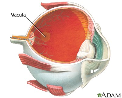
You are born with the vitreous humor in place. Should it be removed, by surgery or trauma, you will not grow another. This is in contrast to the aqueous humor, a clear liquid that fills the space between the cornea that protects the eye and the colored part called the iris (called the anterior chamber of the eye – see diagram). The aqueous humor is manufactured by the ciliary body, a muscular structure that gives rise to the iris. From its manufacture point just behind the iris, the fluid circulates through the pupil, fills the space behind the cornea, and exits via a channel  formed by the junction of the iris and the cornea. This evenly balanced system of fluid manufacture, circulation and exit controls the pressure within the whole eye. No such recycling system exists for the vitreous humor – as in Las Vegas, what happens in the vitreous humor stays in the vitreous humor.

eye anatomy

What happens without recycling?

The  collagen structure within the vitreous humor is an airy honey-comb of interconnected collagen fibrils – microscopic fibers cross-linked and held apart by chemical and electrostatic forces. The network is loosely attached at some points to the retina that lines the inside of the back of the eyeball. The spaces in the honeycomb contain a solution of many minerals and polysaccharide molecules (chains of sugars) dissolved in water. The collagen network is, in part, held open by the pressure of the watery solution. With age, the system has mini-collapses of collagen fibrils, resulting in some clumping of the collagen networks. Floaters are the result. Occasional macrophages (white blood cells that clean up debris) float about, but they are a lonely workforce.

When are floaters more than a nuisance?

A sudden increase in the number of floaters, accompanied by transient light flashes, is sometimes indicative of a segment of the vitreous pulling away from an attachment point on the retina. This condition is known as a posterior vitreous detachment. By itself, a posterior detachment is not a serious problem, but occasionally the point of shrinkage also pulls the retina away from the vascular layer underneath it. Now you have a retinal detachment, and your vision either develops a blind spot or the sensation of a curtain pulled over part of the visual field. Such symptoms require immediate ophthalmologic evaluation and treatment to prevent further detachment of the retina.

Treatment

There is no special treatment for floaters but at times, floaters are troublesome enough for an ophthalmologist to attempt to remove the vitreous humor entirely. The surgery is very difficult and fraught with hazards such as retinal detachment or damage resulting in partial blindness.  Called a vitrectomy, it is a procedure done more often  for other reasons such as  eye trauma. After vitrectomy, the vitreous has to be replaced to maintain the shape of the eye. Research by physicists and bioengineers on suitable replacement substances has been the biggest source of information about the physical nature of the vitreous humor and the origin of the near universal phenomenon of floaters. However, to date the replacement is still done with saline, which is then naturally replaced by the same fluid that fills the anterior chamber.

Natural history of floaters 

What happens once you begin to notice floaters? They come and they go – eventually sinking out of view. The process is very slow because there is no circulation pattern in the vitreous humor and the body has not assigned a vigorous cleanup crew to the problem. Because of this slowness, and because the vitreous passively absorbs substances from the bloodstream via the blood vessels in the retinal layer, coroners sometimes use the vitreous humor to search for toxic substances like drugs at autopsy. Chemical traces remain there after they have disappeared from other body fluids; and. the vitreous humor also retains its integrity longer than other parts of the body.

Plato’s cave

As for “seeing” floaters? You are not actually looking at the clumps of collagen. Just as in Plato’s story about people in a cave interpreting shadows created by firelight as reality, you see only the shadows of floaters cast on the retina by light coming through the pupil. For this reason, floaters are clearest when you are looking at a bright background such as snow or water. Even when you see floaters with your eyes closed, light is passing through the thin eyelid and into the pupil, the only opening in an otherwise light proof box. Who knew these little annoyances could illustrate a philosophy lesson?


Why We Cry..and How We Make the Tears


“It is such a secret place, the land of tears.” The Little Prince, Antoine de Saint-Exupery

Do animals cry? Probably not. Indian gamekeepers told Charles Darwin stories of elephants that shed tears of sadness, and dog lovers have tales of canine tears, but the emotional tears of humans are unparalleled in the animal kingdom.

We are always making tears

All land dwelling animals, including people, make tears constantly. Eyes are windows on the world, and baseline tears are constant window washers. The window pane is the cornea, a thin panel of collagen, containing very few cells, no blood vessels, and more nerves per square inch than any other part of the body. These nerves signal alarm and summon an army of reflexive tears in response to a speck of dust, a cold wind, or a whiff of an onion. Reflexive tears, which are just a lot of baseline tears, wash out intruders and fill in dry patches on the cornea, keeping it clear and moist to focus light entering the eye. Emotional tears appear in humans during infancy, but not immediately. The crying that infants first do to signal their needs is much like the crying of little chimpanzees – tearless. Emotional tears come later, just like talking. Both are outward expressions of the lives of our minds, and they take a while to learn.

What tears are made of

Tears are much more than little beads of salt water running down your face. They are a three layer sandwich. The oil-containing molecules in the outer layer tighten up the surface of the watery middle layer to keep it from spilling over eyelid and sliding off the surface it protects. The oil floats on the watery middle layer and smooths its surface, optimizing the passage of light through to the eye’s interior. The third part of the sandwich, inside and closest to the eye, is the mucinous layer, kind of a gluey protein that helps tears stick to the eyeball. The mucinous proteins capture and kill biologic intruders like bacteria and viruses, and soak up some of the watery layer to help transfer nutrients, oxygen and moisture to the cornea. Both the oil and mucin slow evaporation of tears as blinking spreads them over the eye.

Evaporation and drainage

Dry spots appear on the cornea after just fifteen non-blinking seconds – easy to do while concentrating or daydreaming. Even with blinking, tears evaporate, or they drain out from the eye into the nose via two tiny lacrimal ducts on the upper and lower eyelids near the nose. If these ducts become scarred or blocked by infection, tears overflow. Six to 10% of babies are born with tear ducts not yet open, but 95% of these will open by age one without any attempt at surgical repair. Conversely, one way of treating dry eyes is to block these ducts with small plastic pellets.

Dry eyes

Too few tears, tears with abnormal composition, and decreased blinking cause dry eyes that itch, sting, burn, get red, and cause blurred vision. Dry eyes are an increasing problem in our air-conditioned, airline-traveling, contact lens-wearing, Lasiked, medicated and aging society. The list of drugs that dry eyes includes many commonly prescribed classes: decongestants, antidepressants, antihypertensives, antihistamines, beta-blockers, hormones, diuretics, ulcer medications, acne drugs, and oral contraceptives. Other causes of dry eyes are infections and immune diseases like rheumatoid arthritis and Sjogren’s syndrome as well as  radiation and radioactive iodine treatment can also damage the tear producing cells. All of these conditions damage the tear producing cells  – the machinery for tear production. 

Remedies for dry eyes 

Treatment of dry eyes is always aimed at removing offending problems and increasing lubrication. The usual tactics include artificial tears, wind protection, air humidification, cessation of unnecessary medications, and treatment of underlying diseases and infections. Excessive tearing often means allergies, or blocked lacrimal ducts leading to poor drainage of baseline tears into the nose.


The tear producing machinery

The medical conditions mentioned above shut down tear production and cause pain and swelling in the lacrimal gland, a spongy little structure tucked up under the upper outer corner of the eyelid. The lacrimal gland is the tear producing factory, aided by the oil-producing Meibomiam glands near the eyelashes, and a cluster of mucin-producing cells in the eyelid lining.

The controls for the machinery

When the lacrimal glands get a call for more tears, either reflexive or emotional, the messages come through the autonomic nervous system, which oversees the automatic functions of the body. Reflexive tears spring from messages sent from the eye and nose. Emotional tears come from messages sent by the limbic system, the deepest and oldest part of the brain, the part that conjures up feelings.

What are emotional tears?

What are emotional tears? Are they just more voluminous baseline tears? Or does emotional crying rid us of “humors of the brain,” as Hippocrates thought? In Roman times, mourners used small glass vials called lachrymators to collect their tears for burial with the one for whom they cried. In today’s laboratory, emotional tears are almost as hard to come by as research money to investigate them. Some dedicated men such as Professor William H. Frey II (Dept. of Pharmaceutics at the U. Of Minnesota) have learned enough to suggest that tears of grief rid the body of some of the products of stress, supporting the claim that crying makes people feel better. Compared to reflexive tears, emotional tears contain up to 25% more proteins, of classes related to stress. Why? No one knows – yet. Emotional tears are still a land of mystery, part of the unique expression of inner life that separates the human animal from the others.


What is Macular Degeneration?

eye

To understand age-related macular degeneration (AMD), the most common cause of legal blindness in older people, you need to understand the macula.  First, think of the eye as a hollow globe made of three layers.  The white, outside layer is the sclera. Inside the sclera is a middle layer called the choroid, which contains networks of tiny blood vessels called capillaries. The inner layer, called the retina, contains pigment-filled cells that absorb light, light receptor cells call rods and cones that change light into electrical information, and nerve cells that transmit that information to the brain. In the center of the retina, there is a tiny yellow spot about the diameter of a pencil eraser called the macula lutea (Latin for yellow spot).

What does the macula do? Try looking at a star to find out.

The macula contains only cone receptors, which detect color, and is responsible for sharply focused vision. If the macula degenerates, visual clarity is lost. The other 96% of the retina contains mostly rods, non-color receptors that perceive dim light and movement and are responsible for peripheral vision.  When you look at stars, you see them only with your peripheral vision. Trying to focus on a single star makes it disappear because the macular cones require more light than the night sky offers.  Imagine the disappearance of anything you try to focus on, in any kind of light, and you have some grasp of what macular degeneration is like.

When the macula degenerates

Age-related macular degeneration moves slowly, with many years between the first visible changes in the retina and the onset of symptoms. The first symptoms come from decreasing visual sharpness. Reading speed declines. Reading glasses no longer work for fine print. Road signs look blurred. With progression, dark patches or blank spots appear when patients focus on faces or print, and sometimes straight lines appear wavy. Legal blindness (best corrected vision less than 20/200) comes later and is never total because peripheral vision is spared, even though some of the same degenerative changes affect other parts of the retina. The macula is simply so tiny and so specialized that small areas of damage interfere with the ability to see clearly.

What’s the cause of macular degeneration?

No one knows for certain what causes macular degeneration but ophthalmologists suspect that the vascular anatomy of the retina plays a significant role. While the macula looks yellow, the rest of the retina has a reddish hue because of an overlying bed of capillaries, branches of the retinal arteries and veins which spread like a tree over the retinal surface. The tree stops at the macula and this vital region depends solely on blood flow through the network of tiny capillaries in the choroid layer for oxygen and nutrients and for clearing away the products of its high rate of energy use. The first changes that signal possible AMD, visible long before any symptoms appear, occur in the microscopic space between the choroid layer and the retina and are called Drüsen.

Signs of deterioration

Drüsen are yellowish accumulations of fats and proteins and inflammatory substances.  Small, sharper edged patches called hard drüsen are commonly seen in other parts of the retina and do not interfere with vision, but larger, fluffy patches, especially in the area of the macula warrant more frequent follow-up exams because people who have them sometimes, but not always, develop symptoms of AMD.  If visual symptoms occur and drüsen are present, the diagnosis is dry macular degeneration. In about 10-20% of cases of dry macular degeneration, new tiny blood vessels poke through to the retinal layer from the choroid, a process called neovascularization. When this happens, the diagnosis becomes wet macular degeneration which reflects the tendency of these blood vessels to leak and bleed, causing more cell destruction and separation of the retina form its underlying supporting layers.

Who’s at risk for macular degeneration?

The primary risk factors for macular degeneration are age, smoking and increased body mass index. A family history of macular degeneration also increases the chances of its occurrence, but so many different genes are involved that predictive genetic testing is not useful. Diabetes and vascular disease can accelerate the degenerative process. On the positive side, diets rich in green leafy vegetables, particularly those containing abundant antioxidants called carotenoids, as well as diets rich in Vitamins C and E, and those rich in zinc have been associated with lower frequencies of macular degeneration.

Other eye problems are more common

Fortunately, AMD is much less common than other age-related visual problems like presbyopia (poor near vision) and cataracts, both of which are eminently treatable. AMD affects about 2% of people in their 70s. Prevalence jumps to about 14% in Caucasians who reach their 90s, but remains at about 2% in other races. Legal blindness typically does not occur until the eighties.

Treatment

There is no treatment for dry macular degeneration.  The positive correlation between dietary antioxidant consumption and lower rates of AMD has led to the development of the “eye vitamins,” and while studies have shown some slowing of the degenerative process when these vitamins are consumed, there is no evidence that taking them prevents the onset of the disease.

The first line of treatment for wet macular degeneration is injection, directly into the eye, of drugs which block new blood vessel formation. This slows the disease process, but does not cure it. Another treatment is photocoagulation, or the injection of drugs which, once activated by light aimed at the new blood vessels causes them to shrivel.  Laser treatment of troublesome blood vessels is commonly done, but it destroys the retina in the area treated, so its aim is prevention of new problems.  Surgery is sometimes required to drain fluid accumulations or reattach retinal tears.

Preliminary studies suggest that high dose cholesterol lowering drugs (statins) may shrink drüsen, but one worrisome study of data from a large managed care group suggested that statin use for over a year increased rates of progression of dry AMD to wet AMD. Long term prospective studies are needed, and eye exams should be routine for anyone taking statins.

Useful Macular Degeneration websites

https://nei.nih.gov/health/maculardegen/armd_facts

http://macularhope.org

http://www.aao.org/eye-care-for-older-adults

http://www.amd.org/

No more posts.